
Google Research embarks on effort to map a mouse brain
September 26, 2023
Posted by Michał Januszewski, Research Scientist, Google Research

The human brain is perhaps the most computationally complex machine in existence, consisting of networks of billions of cells. Researchers currently don’t understand the full picture of how glitches in its network machinery contribute to mental illnesses and other diseases, such as dementia. However, the emerging connectomics field, which aims to precisely map the connections between every cell in the brain, could help solve that problem. While maps have only been created for simpler organisms, technological advances for mapping even larger brains can enable us to understand how the human brain works, and how to treat brain diseases.
Today, we're excited to announce that the Connectomics team at Google Research and our collaborators are launching a $33 million project to expand the frontiers of connectomics over the next five years. Supported by the Brain Research Through Advancing Innovative Neurotechnologies (BRAIN) Initiative at the National Institutes of Health (NIH) and led by researchers at Harvard University, we'll be working alongside a multidisciplinary team of experts from the Allen Institute, MIT, Cambridge University, Princeton University and Johns Hopkins University, with advisers from HHMI’s Janelia Research Campus. Our project goal is to tackle an immense challenge in neuroscience: mapping a tiny fraction (2-3%) of the mouse brain. We will specifically target the hippocampal region, which is responsible for encoding memories, attention and spatial navigation. This project is one of 11 funded by the NIH's $150 million BRAIN Initiative Connectivity Across Scales (BRAIN CONNECTS) program. Google Research is contributing computational and analytical resources to this effort, and will not receive any funding from the NIH. Our project asks a critical question: Can we scale and speed up our technologies enough to map the whole connectome of a mouse brain?
The modern era of connectomics
This effort to map the connectome of a small part of the mouse brain builds on a decade of innovation in the field, including many advances initiated by the Connectomics team at Google Research. We hope to accomplish something similar to the early days of the Human Genome Project, when scientists worked for years to sequence a small portion of the human genome as they refined technologies that would enable them to complete the rest of the genome.
In 2021, we and collaborators at Harvard successfully mapped one cubic millimeter of the human brain, which we released as the H01 dataset, a resource for studying the human brain and scaling connectomics technologies. But mapping the entire human brain connectome would require gathering and analyzing as much as a zettabyte of data (one billion terabytes), which is beyond the current capabilities of existing technologies.
Analyzing a mouse connectome is the next best thing. It is small enough to be technically feasible and could potentially deliver insights relevant to our own minds; neuroscientists already use mice to study human brain function and dysfunction. By working together to map 10–15 cubic mm of the mouse brain, we hope to develop new approaches that will allow us to map the entire remainder of the mouse brain, and the human brain thereafter.
 |
| Neuroscientists have been working for decades to map increasingly larger and more complicated connectomes. |
One of biology’s largest datasets
In this connectomics project, we will map the connectome of the hippocampal formation of the mouse brain, which converts short-term memories into long-term memories and helps the mouse navigate in space. The mouse hippocampal formation is the largest area of any brain we’ve attempted to understand in this way. Through mapping this region of the mouse brain, we will create one of the largest datasets in biology, combining about 25,000 terabytes, or 25 petabytes of brain data. For reference, there are about 250 billion stars in our Milky Way Galaxy. If each of those stars was a single byte, it would take 100,000 Milky Way Galaxies to match the 25 petabytes of data that the project will collect when mapping a small region of the mouse brain.
To illustrate the hippocampal project’s scale, we calculated the number of Pixel phones (shown as stacks of Pixels below) needed to store the image data from the completed connectome projects that mapped the roundworm and fruit fly brains, as well as for the mouse hippocampal region and entire mouse brain projects, which are just getting started.
Then, we compared the heights of each Pixel stack to familiar objects and landmarks. It would take a stack of 100 Pixels, as tall as a four-year-old girl, to store the image data for the fruit fly brain, the largest completed project thus far. In contrast, the mouse hippocampal connectome effort will require storage equivalent to more than 48,800 Pixels, reaching as high as the Empire State Building. The animation below shows how the mouse hippocampal project will surpass the scale of previous connectome projects.
 |
| We are partnering with several collaborators to build a connectome (a map of the connections between brain cells) for the hippocampal region of a mouse brain. This project will create the largest connectomic dataset ever, surpassing the scale of previous projects that mapped the smaller roundworm and fruit fly brains. We hope this effort will lead to the development of new approaches that will allow us to later map an entire mouse brain. This animation shows how the field of connectomics is scaling up by calculating the number of Pixel phones needed to store the data from various projects. It would take just two Pixels, the height of an olive, to store the roundworm connectome data, while it would take a stack of Pixels the size of Mount Everest to store the data from an entire mouse connectome. |
Understanding the connectome of the mouse hippocampal formation could help illuminate the way our own brains work. For instance, we may find common features between this circuitry in the mouse brain and human brains that explain how we know where we are, how our brains associate memories with specific locations, and what goes wrong in people who can’t properly form new spatial memories.
Opening the petabyte pipeline
Over the last decade, our team has worked to develop tools for managing massive connectomic datasets, and extracting scientific value from them. But a mouse brain has 1,000 times more neurons than the brain of the Drosophila fruit fly, an organism for which we helped build a connectome for a large part of the brain. Starting the mouse brain connectome will challenge us to improve existing technologies to enable us to map more data faster than ever before.
We’ll continue to refine our flood-filling networks, which use deep learning to trace, or “segment”, each neuron’s path through three-dimensional brain volumes made from electron microscope data. We’ll also extend the capabilities of our self-supervised learning technology, SegCLR, which allows us to automatically extract key insights from segmented volumes, such as identifying cell type (e.g., pyramidal neuron, basket neuron, etc.) and parts of each neuron (e.g., axon, dendrite, etc.).
 |
| A flood filling network traces a neuron through three-dimensional brain space. |
We will also continue to enhance the scalability and performance of our core connectomics infrastructure, such as TensorStore for storage and Neuroglancer for visualization, in order to enable all of our computational pipelines and human analysis workflows to operate at these new scales of data. We’re eager to get to work to discover what peering into a mouse’s mind might tell us about our own.
Acknowledgements
The mouse connectomics project described in this blog post will be supported in part by the NIH BRAIN Initiative under award number 1UM1NS132250. Google Research is contributing computational and analytical resources to the mouse connectome project, and will not receive funding from the NIH. Many people were involved in the development of the technologies that make this project possible. We thank our long-term academic collaborators in the Lichtman Lab (Harvard University), HHMI Janelia, and the Denk Lab (Max Planck Institute for Biological Intelligence), and acknowledge core contributions from the Connectomics Team at Google. We also thank John Guilyard for creating the illustrative animation in this post, and Elise Kleeman, and Erika Check Hayden for their support. Thanks to Lizzie Dorfman, Michael Brenner, Jay Yagnik and Jeff Dean for their support, coordination and leadership.
Other posts of interest
-
April 23, 2024
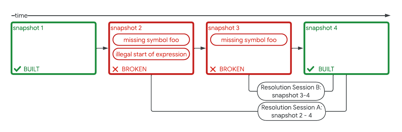
Safely repairing broken builds with ML- Machine Intelligence ·
- Software Systems & Engineering
-
April 17, 2024
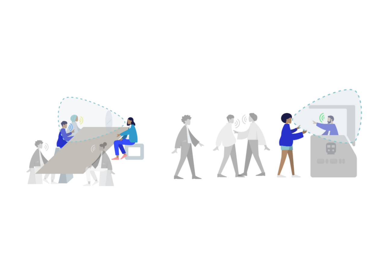
Robust speech recognition in AR through infinite virtual rooms with acoustic modeling- Human-Computer Interaction and Visualization ·
- Machine Perception ·
- Speech Processing
-
April 12, 2024
Contrastive neural audio separation- Machine Intelligence ·
- Sound & Accoustics